Sat, May 4, 2024
Volume 7, Issue 2 (6-2018)
2018, 7(2): 63-68 |
Back to browse issues page


Download citation:
BibTeX | RIS | EndNote | Medlars | ProCite | Reference Manager | RefWorks
Send citation to:


BibTeX | RIS | EndNote | Medlars | ProCite | Reference Manager | RefWorks
Send citation to:
Jafari K, Rezaei A, Samadi V, Hekmatfar S. The Association Between Dental Anomalies and Hypodontia Among 9-20 Years Old Individuals in Ardabil City, Iran: A Causal-Comparative Study. Journal title 2018; 7 (2) :63-68
URL: http://3dj.gums.ac.ir/article-1-317-en.html
URL: http://3dj.gums.ac.ir/article-1-317-en.html


1- Assistant Professor, Department of Prosthodontics, School of Dentistry, Ardabil University of Medical Sciences, Ardabil, Iran.
2- Dental Student, Students Research Committee, Faculty of Dentistry, Ardabil University of Medical Sciences, Ardabil, Iran.
3- Assistant Professor, Department of Pediatric Dentistry, School of Dentistry, Ardabil University of Medical Sciences, Ardabil, Iran.
2- Dental Student, Students Research Committee, Faculty of Dentistry, Ardabil University of Medical Sciences, Ardabil, Iran.
3- Assistant Professor, Department of Pediatric Dentistry, School of Dentistry, Ardabil University of Medical Sciences, Ardabil, Iran.
Full-Text [PDF 605 kb]
(836 Downloads)
| Abstract (HTML) (2393 Views)

Full-Text: (788 Views)
1. Introduction
Hypodontia refers to the developmental absence of at least one deciduous or permanent tooth, excluding the third molars. Hypodontia is one of the most common human dental developmental anomalies [1, 2]. This anomaly causes not only esthetic and physiologic problems but also malocclusion and functional disorders [3]. The prevalence of hypodontia in the permanent dentition has been estimated between 0.15% and 16.2%, depending on the studied population (excluding the third molars) [2, 4, 5]. Furthermore, females were reported to have a higher prevalence of hypodontia [6]. In the primary dentition, hypodontia is not frequent and reported between 0.1% and 2.4% [7].
Hypodontia can result from environmental or genetic factors [2, 8]. Recent advancement in the investigations regarding the genetics of hypodontia has suggested that specific genes, including PAX9 and MSX1, are associated with this anomaly. These genetic factors can also be related to the delayed dental development of the remaining teeth [9-11].
Some studies have indicated that hypodontia is frequently associated with other dental anomalies, such as peg-shaped lateral incisors, transposition, impaction, taurodontism, ectopic eruption, retained deciduous teeth, and enamel hypoplasia. According to previous studies, the development of permanent teeth in children with dental agenesis was similar to children with normal dental development [12]. However, few studies have investigated whether any relationship exists between hypodontia and dental anomaly [13]. In this regard, there is an inconsistency in the study findings due to differences in the investigated population, gender distribution, and age range of the subjects. Accordingly, this paper aimed to analyze the radiographic findings of the tooth anomalies in patients with hypodontia.
2. Materials and Methods
This causal-comparative study was approved by the Ethics Committee of Ardabil Medical University, Iran. This cross-sectional study was a retrospective survey of dental panoramic radiographs of patients referring to the Ardabil radiology centers during 2016-2017.
The study radiographs belonged to 101 subjects whose 1-4 permanent teeth were congenitally absent, except for the third molars. The age range of the subjects was 9-20 years. These subjects were compared to 182 cases in the control group with full dentitions under the same condition. The patients with the history of systemic diseases, any syndromes (i.e. Down syndrome, cleidocranial dysostosis, and ectodermal dysplasia), cleft lip, cleft palate, trauma or fracture of jaws, orthodontic treatment, and the radiographs with low quality were excluded from this study.
Panoramic radiographs were used to investigate the presence of the following anomalies: 1. Microdontia of the maxillary lateral incisors; 2. Retained deciduous molars; 3. Impaction; 4. Supernumerary teeth; 5. Infraocclusion of the deciduous molars; 6. Transposition; and 7. Ectopic eruption of unerupted first molars. Panoramic radiographs were assessed, and two investigators examined the records and diagnosed the dental anomalies.
The collected data were analyzed to determine if there was any association between hypodontia and other dental anomalies. Statistical analysis was performed in SPSS (version 16.0; IBM, Armonk, NY). The Chi-squared test was employed to compare the prevalence of dental anomalies associated with tooth agenesis at the significance level of 0.05 (P<0.05). The Odds Ratio (OR) was calculated at 95% Confidence Interval (CI) to assess the strength of the association between tooth agenesis and other dental anomalies. Descriptive statistics and frequency tables were then created for general descriptions of the collected results.
3. Results
Dental panoramic radiographs of 101 subjects (46 males and 55 females) were evaluated carefully. The commonest anomalies in all investigated radiographs were dilacerations, impaction, and retained teeth, in the descending order. Furthermore, 56.6% of teeth agenesis were observed in the maxilla and 43.4% in the mandible. There was no statistical difference between maxilla and mandible with regard to tooth agenesis (P=0.08). The findings revealed that 1 tooth and 4 teeth agenesis were observed in 52.5% and 9.9% of evaluated radiographs, respectively. Table 1 presents the frequency and percentages of the observed anomalies in both groups.
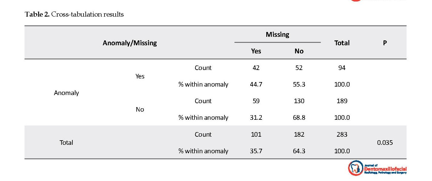
The results of this study showed a significant statistical difference between the groups with and without tooth regarding agenesis (P=0.035) (Table 2). In the group with tooth agenesis, the prevalence of another dental anomaly was lower (P=0.026) with the OR of 1.78. However, there was a higher rate of dental anomalies in the control group.
The highest frequency belonged to the agenesis of mandibular second premolar, then maxillary second premolar, and finally maxillary lateral incisors. The most common anomalies associated with mandibular second premolar and maxillary second premolar were retained teeth and dilacerations, respectively. Furthermore, the most frequently observed anomalies in maxilla and mandible were dilacerations and retained teeth, respectively.
4. Discussion
Previous retrospective studies which investigated tooth agenesis, except for the third molars, reported the prevalence of 20.2% among patients in Ardabil City, Iran [14]. This study aimed to investigate the null hypothesis that there was no association between tooth agenesis and other dental anomalies in children by comparing them with an age- and sex-matched control group. The obtained results of this study did not reveal any significant association between tooth agenesis and other dental anomalies. Therefore, there was no association between the severity of hypodontia and the number of dental anomalies.
In this study, however, the prevalence of teeth anomaly was higher in the control group. The most common dental anomaly associated with hypodontia was retained deciduous molars, affecting 29.5% (n=52) of the total sample (n=176). Likewise, studies conducted by Gomes et al. and Al-Abdullah et al. [15, 16] indicated that the retained deciduous teeth were the most frequent dental anomaly, which affected 30.3% and 29.5% of their hypodontia cases. Furthermore, it was found that retained deciduous molars were more significantly associated with mandibular hypodontia (60.0%), compared to maxillary hypodontia (9.4%). This finding was expected since the most common agenesis in the mandibular hypodontia group was the second premolar, and the retention of mandibular deciduous second molars was prevalent with a long life expectancy [17, 18].
There was also a report of the relationship between hypodontia and impaction [19, 20]. More specifically, this involves the absence or reduced size of the maxillary lateral incisor. The maxillary canine is guided in eruption by the lateral incisor root. If the lateral incisor root is absent or diminutive, eruption guidance is lost and the canine can impact a palatal position. It has been reported that the absence of the lateral incisor increases the prevalence of maxillary canine impaction. One study reported no difference related to the severity of dental agenesis and prevalence of displaced maxillary canine [21].
The limitations of this study were the small sample size and overlooking the genetic influences on the concomitant occurrence of dental anomalies. Also, the panoramic radiographs were the only diagnostic tools. It is recommended to conduct future genetic research studies in this topic.
5. Conclusion
The prevalence of dental anomalies in the agenesis group was relatively low and evenly distributed among genders and jaws. The most commonly affected teeth were the mandibular second premolar, then maxillary second premolar, and finally maxillary lateral incisors. According to the results of this study, the clinician should be aware of the possibility of associated anomalies in all patients and the accompanying clinical problems. The early recognition and careful management of dental anomalies can prevent prosthodontic or orthodontic treatment complications.
Ethical Considerations
Compliance with ethical guidelines
This study was approved by the Ethics Research Committee of Ardabil Medical University, Iran.
Funding
This research is self-funded.
Authors contributions
Conceptualization: Somayeh Hekmatfar; Methodology: Somayeh hekmatfar, Karim Jafari; Investigation: Somayeh Hekmatfar, Amir Rezaei, Vahid Samadi; Writing: Karim Jafari, Somayeh Hekmatfar; Supervision: Karim Jafari, Vahid Samadi.
Conflict of interest
The authors declared no conflict of interest.
Acknowledgements
The authors would like to thank the Heads of Student Research Committee, Dental Faculty, and Ardabil University of Medical Sciences, for their support.
References
Goya HA, Tanaka S, Maeda T, Akimoto Y. An orthopantomographic study of hypodontia in permanent teeth of Japanese pediatric patients. Journal of Oral Science. 2008; 50(2):143-50. [DOI:10.2334/josnusd.50.143] [PMID]
De Coster PJ, Marks LA, Martens LC, Huysseune A. Dental agenesis: Genetic and clinical perspectives. Journal of Oral Pathology & Medicine. 2009; 38(1):1-17. [DOI:10.1111/j.1600-0714.2008.00699.x] [PMID]
Kokich VG, Kokich VO. Congenitally missing mandibular second premolars: Clinical options. American Journal of Orthodontics and Dentofacial Orthopedics. 2006; 130(4):437-44. [DOI:10.1016/j.ajodo.2006.05.025] [PMID]
Heuberer S, Ulm C, Zechner W, Laky B, Watzak G. Patterns of congenitally missing teeth of non-syndromic and syndromic patients treated at a single-center over the past thirty years, Archives of Oral Biology. 2019; 98:140-7. [DOI:10.1016/j.archoralbio.2018.11.018] [PMID]
Behr M, Proff P, Leitzmann M, Pretzel M, Handel G, Schmalz G, et al. Survey of congenitally missing teeth in orthodontic patients in Eastern Bavaria. European Journal of Orthodontics. 2011; 33(1):32-6. [DOI:10.1093/ejo/cjq021] [PMID]
Endo T, Ozoe R, Kubota M, Akiyama M, Shimooka S. A survey of hypodontia in Japanese orthodontic patients. American Journal of Orthodontics and Dentofacial Orthopedics. 2006; 129(1):29-35. [DOI:10.1016/j.ajodo.2004.09.024] [PMID]
Rakhshan V. Congenitally missing teeth (hypodontia): A review of the literature concerning the etiology, prevalence, risk factors, patterns and treatment. Dental Research Journal. 2015; 12(1):1-13. [DOI:10.4103/1735-3327.150286]
Nieminen P. Genetic basis of tooth agenesis. Journal of Experimental Zoology Part B Molecular and Developmental Evolution. 2009; 312B(Special Issue):320-42. [DOI:10.1002/jez.b.21277] [PMID]
Dhanrajani PJ. Hypodontia: Etiology, clinical features, and management. Quintessence International. 2002; 33(4):294-302. [PMID]
Cobourne MT. Familial human hypodontia-is it all in the gene? British Dental Journal. 2007; 203(4):203-8. [DOI:10.1038/bdj.2007.732] [PMID]
Hekmatfar S, Hoseini S, Mikaili H, Jafari K. Radiographic evaluation of dental development in patients with tooth agenesis. Journal of Dentomaxillofacial Radiology, Pathology and Surgery. 2017; 6(4):123-8.
Matalova E, Fleischmannova J, Sharpe PT, Tucker AS. Tooth agenesis: From molecular genetics to molecular dentistry. Journal of Dental Research. 2008; 87(7):617-23. [DOI:10.1177/154405910808700715] [PMID]
Amini F, Rakhshan V, Babaei P. Prevalence and pattern of hypodontia in the permanent dentition of 3374 Iranian orthodontic patients. Dental Research Journal. 2012; 9(3):245-50. [PMID] [PMCID]
Hekmatfar S, Bagheri A, Jafari K, Zarei S, Heidarzade Z. Incidence of dental developmental anomalies in permanent dentition among Ardabil population, Iran, in 2015-2016. Journal of Oral Health and Oral Epidemiology. 2018; 7(2):64-8.
Gomes RR, da Fonseca JA, Paula LM, Faber J, Acevedo AC. Prevalence of hypodontia in orthodontic patients in Brasilia, Brazil. European Journal of Orthodontics. 2010; 32(3):302-6. [DOI:10.1093/ejo/cjp107] [PMID]
Al-Abdallah M, AlHadidi A, Hammad M, Al-Ahmad H, Saleh R. Prevalence and distribution of dental anomalies: A comparison between maxillary and mandibular tooth agenesis. American Journal of Orthodontics and Dentofacial Orthopedics. 2015; 148(5):793-8. [DOI:10.1016/j.ajodo.2015.05.024] [PMID]
Bjerklin K, Bennett J. The long-term survival of lower second primary molars in subjects with agenesis of the premolars. European Journal of Orthodontics. 2000; 22(3):245-55. [DOI:10.1093/ejo/22.3.245] [PMID]
Bjerklin K, Al-Najjar M, Karestedt H, Andren A. Agenesis of mandibular second premolars with retained primary molars: A longitudinal radiographic study of 99 subjects from 12 years of age to adulthood. European Journal of Orthodontics. 2008; 30(3):254-61. [DOI:10.1093/ejo/cjn027] [PMID]
Mossey P, Campell HM, Luffingham JK. The palatal canine and the adjacent lateral incisor: A study of a west of Scotland population. British Journal of Orthodontics. 1994; 21(2):169-74. [DOI:10.1179/bjo.21.2.169]
Peck S, Peck L, Kataja M. Site-specificity of tooth agenesis in subjects with maxillary canine malpositions. The Angle Orthodontist. 1996; 66(6):473-6. [PMID]
Laganà G, Venza N, Lione R. Associations between tooth agenesis and displaced maxillary canines: A cross-sectional radiographic study. Progress in Orthodontics. 2018; 19(23):2-6.
Received: 2018/01/2 | Accepted: 2018/04/28 | Published: 2018/06/1
| Rights and permissions | |
 | This work is licensed under a Creative Commons Attribution-NonCommercial 4.0 International License. |
Articles Copyright © The Author(s).
Owned by Guilan University of Medical Sciences.
Co-published by Negah Institute for Scientific Communication.
Contact Information
The end of professor Samii Blv, Guilan University of Medical Sciences Complex, Dental School, Rasht, Iran.
Journal Tel : +9813 33486428
Publisher Tel : +9821 86037228 , 86036497
Email: den3djournal@gums.ac.ir
