

BibTeX | RIS | EndNote | Medlars | ProCite | Reference Manager | RefWorks
Send citation to:
URL: http://3dj.gums.ac.ir/article-1-224-en.html


2- oral and maxillofacial Radiology, Qazvin University of Medical Sciences, Qazvin, Iran
3- Private practice
4- Dental School, Alborz University of Medical Sciences, Karaj, Iran
Iranian Dental Students’ Level of Knowledge Regarding the Interpretation of Radiographic Images of Oral Lesions: Is the Standard Training Sufficient?
Abstrat
Introdouction:
Dentists need to be able to diagnose jaw lesions due to professional responsibilities to refer affected patients for expeditious treatment as needed. The objective of this study was to assess the knowledge of senior dental students in Qazvin University of Medical Sciences regarding the interpretation of radiographic images of oral lesions. The study took place in 2011–2012.
Materials and methods:
This descriptive-analytical trial involved 36 dental students enrolled in the practical and theoretical radiology course and oral medicine during their final educational session. A questionnaire was designed, and students were asked to express their diagnoses following observation and interpretations of nine different diagnostic aspects in dental images in 3 questions and write the first probable diagnosis for 10 items in one question. The students’ scores were calculated and statistically analyzed by one-sided analysis of variance and student t-tests.
Results:
The students’ mean score was 14.32 ± 2.09, out of a maximum score of 20. The maximum and minimum scores of participants were 18.0 and 10.5, respectively. No significant differences were noted between the scores of male and female students.
Conclusion:
In total, participating students demonstrated an acceptable level of knowledge in the interpretation of radiographic images.
Key words:
Dental Radiography, Dental Students, Knowledge.
Introdouction
Radiographic evaluation along with clinical examination is necessary for the diagnosis of lesions. Radiography can help clinicians to detect lesions (especially bone lesions), such as primary tumors, before their clinical appearance. Radiography is also used to evaluate the size and extension of lesions and in the development of treatment plans. (1,2,3) However, one of the major limitations of radiography is that it depends on the individual’s skills and ability in interpreting radiographs.
Considering the responsibilities of dental students in providing community oral health, it is essential to be aware of the radiographic interpretation of oral lesions.
If doctors and dentists, who are the main public health providers, have no knowledge about interpretation of radiographic lesions, these lesions can progress to an advanced stage, confronting the patient with dangerous problems.(4) Some research results in the field of knowledge and practice of dentists, doctors, and dental students have shown obvious weaknesses in the area of malignant oral lesions, emphasizing the need to improve learning methods for these
occupational groups.(5,7) The present study assessed the knowledge of the diagnosis and differentiation of jaw lesions among senior dental students at one Iranian university during 2011–2012.
Materials and Methods
Subjects: 36 senior dental students of Qazvin University of Medical Science, Iran, participated in this descriptive-analytical study. They had passed practical and theoretical radiology and oral medicine studies. Technical Information: Two oral medicine specialists and one oral and maxillofacial radiologist designed the questionnaire, based on the textbook Oral Radiology: Principles and Interpretation by Michael J. Pharoah Stuart C. White 2009. They selected the criteria required to interpret radiographic images for lesions (location, local or generalized, unilateral or bilateral, well-defined or ill-defined), forms of lesion (regular,
irregular, or crenate), whether lucent or opaque, internal structure, and effect on surrounding structures. All slide images were selected from referred patients or from published articles, and participants had not seen the images previously. The questionnaire consisted of four questions; the total score for each question was five. Thus, the overall questionnaire was set based on a potential score of 20. In Question 1, various radiological features were shown, and the students were asked to write the first probable diagnosis (one of three related diagnoses was acceptable). The 10 radiological features shown consisted of the following:
ameloblastoma; bone marrow space; odontoma; odontogenic keratocyst (OKC); cementoblastoma; radicular cysts; stafne; traumatic bone cyst (TBC); florid cemento-osseous dysplasia (FLCOD); and incisive canal cyst. The correct answer to each item had 0.5 scores. Question 2 involved the condition of a 22-yearold
woman who had been referred for a routine checkup. During clinical evaluation, a bony hard swelling was detected. Students were asked to write their interpretation and diagnosis based on a dental panoramic Radiographs of this patient ( Figure 1). This question was designed to evaluate knowledge about odontogenic cysts (Dentigerous). The correct answer to 8 items of the lesion (location, local or generalized, unilateral or bilateral, well-defined or ill defined, forms of lesion—regular, irregular or crenate—lucent or opaque, internal structure, the effect on surrounding structures) carried a 0.5 score and differential
diagnosis (up to 3 items) had 1 score (totally 9 different diagnostic aspect). In Question 3, it was explained that a 40-year-old man had come to the clinic with a swelling of the jaw, detected three months earlier, and that the overlying mucosa was normal (Figure 2). ). This question was designed to evaluate knowledge about malignant jaw tumors (Osteosarcoma). Question 4 involved a 35-year-old man who had come to the clinic complaining of swelling and pain and tingling of the jaw (Figure 3). This question was designed to evaluate knowledge about benign jaw tumors (Ameloblastoma). As in Question 2, for Questions 3 and 4 students were asked to write their interpretation and diagnosis based on dental images (panoramic, periapical, occlusal) of the patient. The correct answer to 8 items of lesions (location, local or generalized, unilateral or bilateral, well-defined or ill-defined, forms of lesion—regular, irregular or crenate—lucent or opaque, internal structure, the effect on surrounding structures) had a score of 0.5; differential diagnosis (up to 3 items) had a score of 1. As mentioned above, the maximum score for each question was 5. Images were selected with sufficient resolution. To make responses easier and less time-consuming, questions and answers were located on one page. The Department of Oral Radiology and Oral Medicine of Qazvin University of Medical Sciences confirmed the content validity of the questionnaire, which was also confirmed by three specialists in this field. To increase the external validity (face validity) of the questionnaire, questions were typed in a clear and sufficiently large font. To confirm the reliability of the test, 7 students were evaluated in a pilot study. With test-retest, reliability was confirmed (p = 0.832). Equal and appropriate conditions were established for students to view the slides. Subjects were required to respond in one session at the given time. Statistical Analysis: The measure of central tendency (mean, standard deviation, standard error, and upper and lower limit of confidence interval)
was evaluated. We reported the average score for each question and compared the results. Student t-test and one-way ANOVA was used to compare scores. Scores were analyzed using the statistical software SPSS (Statistical Package for Social Sciences), version 18.0. Significance was set at the 95% level. Ethics: We confirm that the students’ information remained confidential, and that data were anonymized and de-identified prior to analysis.
Results
The average score for all students was equal to 14.32 ± 2.09 (the potential maximum score was 20), or 71.6% ± 10.45%. The maximum score was 18.0 and the minimum score was 10.5, respectively. Breaking the results by question (out of a maximum score of 5 for each) the mean scores for responses were as follows respectively: Question 2 (diagnosis of odontogenic cyst): 3.78 ± 0.57; Question 4 (diagnosis of benign jaw tumor): 3.69 ± 0.64; Question 1 (net diagnosis of lesions): 3.53 ± 1.11; Question 3 (diagnosis of malignant jaw tumor): 3.31 ± 0.92. There was no significant difference in responses between questions. Table 1 presents the scores of students for Questions 1 to 4. In addition, there was no significant difference in responses between males and females. Table 2 presents the scores of male and female students for each question and their total scores.
Discussion
This study evaluated the knowledge of senior dental students of Qazvin University of Medical Science (Iran) regarding a range of radiological mfeatures. 10 features in question 1 evaluated, and in question 2-4, nine aspect of each lesion were evaluated. ameloblastoma; bone marrow space; odontoma; odontogenic keratocyst (OKC); cementoblastoma; radicular cysts; stafne; traumatic bone cyst (TBC); florid cemento-osseous dysplasia (FLCOD); and incisive canal cyst (in
Question 1); dentigerous cyst (in Question 2); osteosarcoma (in Question 3); and ameloblastoma (in Question 4) were evaluated. The results showed that the students had appropriate knowledge of the radiographic interpretation of oral lesions. The average score was 14.32 out of 20. The participants had acceptable
knowledge (> 70%) about the characteristics of radiographic images of bony lesions of the jaw. Students had almost equal knowledge about the diagnosis and interpretation of the different jaw lesions, such as cysts and benign and malignant tumors (no statistical differences), which represents adequate training to students in various aspects. Despite numerous studies examining the knowledge, attitude, and practice of physicians, dentists, and dental students about oral cancer, there are few studies that have been conducted in the same way as the present study. In assessing the capability of graduating dental students to interpret panoramic radiography Razmus et al. (1993), concluded that it was important to the detection of lesions and anatomical landmarks, and to the ability to express differential diagnosis for lesions. They showed that factors significantly affecting student performance were the number of lecture hours provided, whether or not students took panoramic radiographs of patients, and the method of student selection as participants.(7) Taheri et al. (2010) assessed knowledge, attitude, and practice of students in Tehran, Iran, in the differential diagnosis of malignant lesions of the jaw from a radiographic aspect. The average scores recorded in this study were less than half the total.(6) Kamburoğlu et al.(2011) evaluated students’ knowledge and attitudes regarding cone beam computed tomography (CBCT).(8) A self-administeredm questionnaire consisting of 11 questions was designed and administered to students at two universities in Turkey. Statistical results showed that only 63.3% of students had heard of CBCT. No differences were found between the responses of male and female participants for any of the questions, a finding also reflected in the present study. However, Kamburoğlu et al. (2011) reported significant differences between the responses of undergraduate and postgraduate students for the majority of questions. This research suggests that efforts should be made to
improve dental students’ knowledge base regarding CBCT, and that the dental school curriculum should be revised. In addition, Lanning et al. (2006) indicated using specific training programs could increase awareness about radiographic interpretation by up to 72%.(9) Lanning et al. (2006) also showed that follow-up instructions and good training programs could improve knowledge about radiographic interpretation by up to 85%.(10) In this study, the average knowledge of students was 71.6% ± 10.45%, which is compatible with the results of Lanning et al. Also, Colella et al. (2008) evaluated knowledge, attitudes, and practices
regarding oral cancer among Italian dentists, and reported that dentists were aware of major risk factors for oral cancer, but that only half of those surveyed knew how to diagnose oral cancer correctly.(11)Spanish dentists’ knowledge and attitudes about oral cancer was evaluated in 2010 by López- et al. They declared that 87.1% of them needed more training.(5) The questions in the present study involved the minimum knowledge expected of senior dental students, as future general dentists, regarding radiographic appearance; difficult questions and vague images were avoided. Since no student achieved the maximum score (20), it seems there are some weaknesses in the knowledge of senior dental students, as future dentists, in the detection and differentiation of oral lesions. Accordingly, current educational programs in Oral Medicine and Oral Radiology may be unable to meet expectations in the field of oral lesions.
To improve the current situation, there is a need for revision of the training curriculum, more practical courses, and continuing education after graduation in the field of oral lesions. To increase the efficiency of the training courses, similar studies should be undertaken to evaluate knowledge, attitudes, and practices regarding.
Conclusion
In this study, participating students demonstrated an acceptable level of knowledge regarding the interpretation of radiographic images of oral lesions (developmental, cyst, benign or malignant). However, further education is required inthis field.
Acknowledgement
This study is completed in cooperation with the Department of Oral Medicine and maxillofacial radiology of Qazvin University of Medical Sciences, Qazvin, Iran.
The authors would like to thank Prof. Marc Tennant of the University of Western Australia for assistance and advice with this project. Conflict of interest: There is no conflict of interest.
Refrences
1.Wenzel A. A review of dentists’ use of digital radiography and caries diagnosis with digital systems. Dento maxillo
fac Radiol. 2006;35(5): 307-314.
2.Versteeg CH, Sanderink GC, van der Stelt PF. Efficacy of digital intra-oral radiography in clinical dentistry. J
Dent.1997;25(3-4):215-224.
3.White SC, Pharaoh MJ. Oral Radiology Principles and Interpretation. 5th Ed. 2009.p.12-25,55-59.
4.Kowalski LP, Franco EL. Lateness of diagnosis of oral and oropharyngeal carcinoma. Factors related to the tumor, the
patient and the health professionals. Oral Oncol Eur J Cancer. 1994;30(3):167-173.
5.Lopez-Jornet P, Camacho-Alonso F, Molina-Minano F. Knowledge and attitudes about oral cancer among dentists in
Spain. J Eval Clin Practice. 2010;16:129-133.
6.Taheri JB, Moshfeghi M, Anbari F, Namazi Z, Bakhtiari S. Knowledge, Opinions and Practice of Last Year Dentistry
Students of Shahid Beheshti University Regarding Diagnosis and Differentiation of Malignant Lesions. Res. J. Med.
Sci. 2010;4 (3): 238-242.
7.Razmus TF, Williamson GF, Van Dis ML. Assessment of the knowledge of graduating American dental students about
the panoramic image. Oral Surg Oral Med Oral Pathol. 1993;76(3):397-402.
8.Kamburoglu K, Kursun S, Akarslan ZZ. Dental students’ knowledge and attitudes towards cone beam computed tomography
in Turkey. Dento maxillo fac Radiol. 2011;40(7):439-43.
9.Lanning SK, Best AM, Temple HJ, Richards PS, Carey A, McCauley LK. Accuracy and consistency of radiographic
interpretation among clinical instructors using two viewing systems. J Dent Educ. 2006;70(2):149-159.
10.Lanning SK, Best AM, Temple HJ, Richards PS, Carey A, McCauley LK. Accuracy and consistency of radiographic
interpretation among clinical instructors in conjunction with a training program. J Dent Educ. 2006;70(5):545-57.
11.Colella G, Maria Gaeta G, Moscariello A, Angelillo IF. Oral cancer and dentists. Knowledge, attitudes, and practices
in Italy. Oral Oncol. 2008;44:393-399.
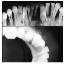
Figure 1. Question 2, panoramic view of a odontogenic cyst in mandible
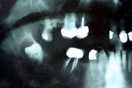
Figure 2. Question 3, Occlusal and periapical view of a malignant tumor in the mandible
Table 1. Knowledge of students about each question separately
|
Question |
N |
Mean |
SD |
SE |
CI 95 % |
Min |
Max |
|
|
Lower |
Upper |
|||||||
|
Q1 |
36 |
3.53 |
1.11 |
0.08 |
3.15 |
3.9 |
1.0 |
5.0 |
|
Q2 |
36 |
3.78 |
0.57 |
0.09 |
3.59 |
3.98 |
2.25 |
5.0 |
|
Q3 |
36 |
3.31 |
0.92 |
0.15 |
2.99 |
3.63 |
1.0 |
5.0 |
|
Q4 |
36 |
3.69 |
0.64 |
0.17 |
3.48 |
3.91 |
2.0 |
5.0 |
Q: Question
SD: standard deviation
Table 2. Knowledge of students about each question in different gender
|
Question |
Gender |
Mean |
SD |
SE |
P value |
|
Question1 |
Male |
4.0 |
0.91 |
0.23 |
0.24 |
|
Female |
3.54 |
1.15 |
0.31 |
||
|
Question2 |
Male |
3.9 |
0.47 |
0.12 |
0.74 |
|
Female |
3.84 |
0.49 |
0.13 |
||
|
Question3 |
Male |
3.33 |
1.05 |
0.27 |
0.66 |
|
Female |
3.50 |
0.96 |
0.26 |
||
|
Question4 |
Male |
3.73 |
0.65 |
0.17 |
0.95 |
|
Female |
3.75 |
0.73 |
0.50 |
||
|
SUM |
F/M |
14.63 |
2.05 |
0.55 |
0.65 |
Received: 2016/09/21 | Accepted: 2016/09/21 | Published: 2016/09/21
| Rights and permissions | |
 | This work is licensed under a Creative Commons Attribution-NonCommercial 4.0 International License. |
Articles Copyright © The Author(s).
Owned by Guilan University of Medical Sciences.
Co-published by Negah Institute for Scientific Communication.
Contact Information
The end of professor Samii Blv, Guilan University of Medical Sciences Complex, Dental School, Rasht, Iran.
Journal Tel : +9813 33486428
Publisher Tel : +9821 86037228 , 86036497
Email: den3djournal@gums.ac.ir
