


BibTeX | RIS | EndNote | Medlars | ProCite | Reference Manager | RefWorks
Send citation to:
URL: http://3dj.gums.ac.ir/article-1-251-en.html


2- Faculty of Dentistry
3- Department of Biostatistics and Epidemiology, Babol University of Medical Sciences, Babol, Iran
The Agreement of Paper and Film Prints in Detection of Dentinal Caries in Panoramic Radiography
Abstract
Introduction:
Dental caries is dramatically common among people. The aim of this study was to evaluate the agreement of paper prints, film prints and monitor in detection of dentinal caries in digital panoramic radiography.
Materials and methods:
In this analytical study, radiographic images of 150 patients referred to a private clinic and who needed panoramic radiographs for various reasons, were used. Images were printed on paper and film. Dentinal caries in molars were classified into three categories; with caries, unknown and without caries. Data were collected in checklist and recorded in SPSS 22. Kappa coefficient was used for obtaining the agreement between the paper prints and film prints and also between monitor and film prints.
Results:
Kappa coefficient between the paper prints and film prints was 0.88 and between monitor and film prints was 0.92.
Conclusion:
According to the results of this study, paper prints could be useful as well as film and monitor in diagnosis of dentinal caries in panoramic radiographies.
Key words:
Dental Caries, X-ray Film, Radiography, Panoramic
Introduction
We encountered different media in digital radiographies; for instance, for displaying (monitor, film prints, etc) and for image storing (CD, film prints, etc).(1) The number of these intermediaries increases the possibility of errors and at the same time reduces the image quality and can lead to many problems in transmission of images.(2)
In recent years, radiologists have tended to use paper prints instead of film prints in digital radiography for storing and recording the images to decrease the costs (3) and to promote environmental health.(4) If there are too many changes and loss of quality of images in recording and storing by paper prints, many problems can be created in diagnosis and the treatment plan and even the function of digital radiography may be questionable.(5) Numerous studies have been done on the comparison of these intermediaries.(6-14) On the other hand, panoramic radiography is widely used for the diagnosis and screening of patients with dental problems; although it is not as useful as periapical radiography for detecting small carious lesions.(15)
The studies have revealed that there is good agreement between panoramic radiography and intraoral in detecting dentinal caries.(16)
Considering all these points, the aim of this study was to evaluate the agreement of paper prints and film prints in the detection of dentinal caries in panoramic radiography.
Materials and Methods
In this analytical study, radiographic images of 150 patients referred to a private clinic and who needed panoramic radiographs for various
reasons, were used. Inclusion criteria for Methodspanoramic radiography were: a) image without technical or positional error; and b) having at least 24 teeth including first and second molars in each quadrant. The present study was approved by the Ethics Committee of Babol University of Medical Sciences (with number of 5121). In this study, all of the panoramic radiographies were obtained by panoramic unit (Cranex D, Tuusula, Finland). Afterwards, they were analyzed in the following three ways:
1) saved on CD and observed on 19-inch monitor LCD Flatron LG E1941 (LG electronics, Seoul, Korea).
2) printed on 160-gram glossy paper (Mondi, Vienna, Austria) by Canon i-sensys LBP 6030B printer (2400 dpi) (Canon, ho chi minh, Vietnam).
3) printed on film by Drypix prima printer (250 dpi) (Fujifilm Corporation, Sendai, Japan).
The digital images were interpreted on the monitor in a darkened environment. Film prints were viewed in a semi-darkened room with light transmitted through view box to film. Paper prints were viewed in normal room conditions. The radiographs were evaluated independently by two observers who were radiologists with at least a 10-years’ experience and their opinions were recorded. In the event of any disagreement, a third radiologist was asked to share his opinion so that one opinion consensually was announced and their agreement was recorded.
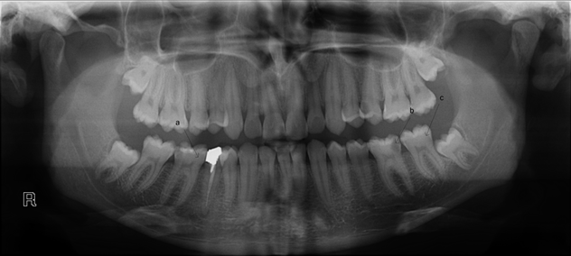
The first and second molars according to presence of dentinal caries in occlusal and proximal surfaces were classified into three groups: 1) with caries; 2) unknown; 3) without caries (Figure 1). Data were collected in checklist and recorded in SPSS 22 Kappa agreement rate of the data was obtained and analyzed.
Results
In the present study, panoramic radiographs of 150 patients (67 males [45%] and 83 females [55%]) aged 15–56 years were used that
included 1,200 teeth with 3,600 dental surfaces; occlusal and proximal surface(mesial, distal).
Table 1 shows the prevalence of dentinal caries according to presence of caries in film prints, paper prints and monitor. Kappa coefficient rate between film prints and paper prints was 0.88 and 0.92 between monitor and film prints, respectively. Table 2 shows the prevalence of dentinal caries according to location of caries in film prints, paper prints and monitor. Paper prints, monitor and film prints were compared according to location of caries. There was high agreement rate between paper prints and film prints with Kappa coefficient rate of 0.90 in the occlusal site and high agreement was observed between monitor and film prints with Kappa coefficient rate of 0.93 in the occlusal site. Kappa coefficient rate was 0.87 between film prints and paper prints and 0.92 between monitor and paper prints in proximal sites, respectively.
Discussion
The results of this study showed that there is a high agreement between film prints and paper prints in detection of dentinal caries, which is compatible to a study by Mehralizadeh et al.(2)They studied the diagnostic efficacy of intraoral paper print of a digital radiograph in detection of proximal dentinal caries on 320 extracted premolars. Their results showed that paper print is almost the same as monitor in terms of diagnostic quality. They found that paper print could be used as a device of digital radiography.(2)
Regarding paper print, there are printers such as ink, laser, and dye sublimation on the market. Among them, the quality of dye sublimation printers is usually higher than the other two; nevertheless, their usage is not economical.(15) However, Schulze et al.(1) and Shafiee et al.(14) concluded that quality of assessment tools such as paper print has been increasing and is quite similar to that of film print in clinical diagnosis by the advancement of technology in the field of oral radiology.(17)
In the present study, the dentinal lesions of the proximal and occlusal surfaces were evaluated. It was concluded that there was no significant difference between paper print and film print. Nevertheless, Bley et al. evaluated the diagnostic quality of radiography on paper prints versus film in high-contrast and low-contrast test objects of the phantom. They stated that paper print, as compared with film print, was not appropriate to diagnose small-sized lesions.(18)
In fact, this difference was due to the lesion size. Since the present study was performed on panoramic radiographs, the diagnostic detection was only on dentinal lesions. Therefore, further studies on periapical radiographs are needed to evaluate smaller lesions. In this study, panoramic radiography was used. Some advantages of this technique include relatively lower dosage of radiation, cost-effectiveness, and production of a single image for both dental arches. Given that, this technique has low accuracy in small lesions. In this paper, dentinal lesions (medium-sized lesions), were evaluated. According to Hosseini et al., panoramic radiography has appropriate accuracy compared with intraoral radiography.(16) In order to achieve these results, panoramic radiographies were compared with paper prints and the results stated that there is a high agreement between them in detection of dentinal caries, but in the case of small lesions, further studies are needed.
Conclusion
According to the results of this study, paper prints, as opposed to film prints, could be useful in diagnosis of dentinal caries in panoramic radiographies.
Acknowledgments
The authors are grateful to Babol University. This study was a part of thesis and research project 683 which was supported and funded byBabol University of Medical Sciences.
References
1.Schulze RK, Schulze D, Voss K, Rottner M, Keller HP, Dollmann K, et al .Quality of individually calibrated customary printers for assessment of typical dental diagnoses on glossy paper prints : a multicenter pilot study Oral Surg , Oral Med, Oral Pathol , Oral Radiol , and Endod.2008; 106(4):578-86.
2. Mehralizadeh S, Sadri. Evaluation of intra oral digital radiography on paper print in proximal dentin caries detection. Journal of Research in Dental Sciences.2009; 6(2):25-33.
3.Fallahzadeh F, Tayyebi A, Tofangchiha M, Modirfallah H, Safarzadeh sh. Agreement of Bitewing and Digital Panoramic Radiographies in the Detection of Proximal Caries. Journal of Kerman University of Medical Sciences. 2013; 20(4):343-53.
4.Cawson R, Odell E, Porter S. Cawson’s essential of oral pathology and oral medicine. 10th edition. New York: Churchill Livingstone, 2015.
5. Schulte AG, Wittchen A, Stachniss V, Jacquet W, Bottenberg P. Approximal caries diagnosis after data import from different digital radiography systems : interobserver agreement and comparison to histological hard-tissue sections.Caries Res.2008; 42(1):57-61.
6.Crawley DA, Longbottom C, Cole BE, Ciesla CM, Arnone D, Wallace VP, et al. Terahertz pulse imaging: a pilot study of potential applications in dentistry. Caries Res.2003; 37(5):352-9.
7.Hintze H. Screening with conventional and digital bite-wing radiography compared to clinical examination alone for caries detection in low-risk children. Caries Res.1993; 27(6):499-504.
8.Nikneshan S, Mashhadi Abbass F, Akbarzadeh Baghban AR, Shahovi L. In vitro carious lesion detection on D, E, and F-speed radiographic films. Journal of Dental School Shahid Beheshti University of Medical Sciences،2010; 28(2):64-70.
9.Lyttkens K, Kirkhorn T, Kehler M, Andersson B, Ebbesen A, Hochbergs P, et al. Evaluation of the image quality of ink-jet printed paper copies of digital chest radiographs as compared with film: a receiver operating characteristic study. J Digit Imaging.1994; 7(2):61-68.
10.Otis LL, Sherman RG. Assessing the accuracy of caries diagnosis via radiograph. Film versus print. J Am Dent Assoc.2005; 136(3):323-30.
11.Tsang A, Sweet D, Wood RE. Potential for fraudulent use of digital radiography. J Am Dent Assoc.1999; 130(9):1325-9.
12.Kirkhorn T, Kehler M, Nilsson J, Lyttkens K, Andersson B, Holmer N-G. Demonstration of digital radiographs by means of ink jet-printed paper copies: pilot study. J Digit Imaging.1992; 5(4):246-51.
13.Abesi F, Mirshekar A, Moudi E, Seyedmajidi M, Haghanifar S, Haghighat N, et al. Diagnostic accuracy of digital and conventional radiography in the detection of non-cavitated approximal dental caries. Iranian J Radiol.2012; 9(1):17-21.
14.Shafiee A, Atala A. Printing Technologies for Medical Applications. Trends in molecular medicine.2016; 22(3):254-65.
15.White SC, Pharoah MJ. Oral Radiology: Principles and Interpretation. 7th ed. Philadelphia: Saunders,2014; chapter 10,P:166.
16.Hoseini Zarch. SH, Javadian Lanaroodi. A, Shafagh Motlagh. M. comparison between two digital panoramic radiography techniques for proximal caries detection. J Dent Mater Tech 2013; 2(2):54-8 .
17.Schulze RK, Grimm S, Schulze D, Voss K, Keller H-P, Wedel M. Diagnostic yield of ink-jet prints from digital radiographs for the assessment of approximal carious lesions: ROC-analysis. Eur J Radiol.2011; 79(2):277-82.
18. Bley TA, Kotter E, Saueressig U, Springer OS, Fisch D, Ghanem NA, et al. Using receiver operating characteristic methodology to evaluate the diagnostic quality of radiography on paper prints versus film. AJR.2003; 181(6):1487-90.
Figure 1.Carious lesions classification: a) unknown; b) with caries; c) without caries
Table1: Prevalence of dentinal caries according to presence of caries in paper prints, film prints and monitor
|
Tools Diagnosis |
Film print N(%) |
Paper print N(%) |
Film print N(%) |
Monitor N(%) |
|
With caries |
420(11.67) |
395(10.97) |
420(11.67) |
398 (11.06) |
|
Unknown |
7(0.19) |
5(0.14) |
7(0.19) |
4(0.11) |
|
Without caries |
3173(88.14) |
3200 (88.89) |
3173(88.14) |
3198(88.83) |
|
Kappa |
0.885 |
0.925 |
||
Table2: Prevalence of dentinal caries according tolocation of caries
|
Position |
occlusal |
proximal |
||||||
|
Film print N(%) |
Monitor N(%) |
Film print N(%) |
Paper print N(%) |
Film print N(%) |
Monitor N(%) |
Film print N(%) |
Paper print N(%) |
|
|
Diagnosis (D) |
118 (9.83) |
112 (9.33) |
118 (9.83) |
115 (9.58) |
302 (12.58) |
286 (11.92) |
302 (12.58) |
285(11.88) |
|
Lack of diagnosis (LD) |
1082 (90.17) |
1088 (90.67) |
1082 (90.17) |
1085 (90.42) |
2098 (87.42) |
2114 (88.08) |
2098 (87.42) |
2120 (88.33) |
|
kappa |
0.933 |
0.900 |
0.922 |
0.879 |
||||
Received: 2017/02/21 | Accepted: 2017/02/21 | Published: 2017/02/21
| Rights and permissions | |
 | This work is licensed under a Creative Commons Attribution-NonCommercial 4.0 International License. |
Articles Copyright © The Author(s).
Owned by Guilan University of Medical Sciences.
Co-published by Negah Institute for Scientific Communication.
Contact Information
The end of professor Samii Blv, Guilan University of Medical Sciences Complex, Dental School, Rasht, Iran.
Journal Tel : +9813 33486428
Publisher Tel : +9821 86037228 , 86036497
Email: den3djournal@gums.ac.ir
